相册共1张
点击图片可以查看相册
STEMdiff™ NK Cell Kit
- 类型:培养基
- 品牌:Stemcell
规格: 1Kit
货号: 100-0170
价格: ¥20276.00
|
该试剂盒可以在无血清和饲养细胞的条件下,使多能干细胞(hPSC),分化成NK细胞。 STEMdiff™ NK细胞试剂盒首先利用无动物源成分的 STEMdiff™ Hematopoietic - EB试剂形成EB,然后进一步分化成CD34。该试剂盒包含EB形成所需的所有试剂:
• StemSpan™ SFEM II |
||||||
|
优点: |
||||||
|
- 一致。使用无血清和无饲养层的配方,消除了因血清和基质细胞系引起的差异。 - 均一。用AggreWell™可以产生均一的EB,降低可变性。 - 产量高。每个PSC衍生的CD34+细胞分别可产生210个CD56 + NK细胞。 - 方便。避免基于培养基质细胞所需的额外传代步骤。 |
||||||
|
数据: |
||||||
|
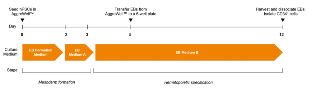
图1.STEMdiff™造血-EB祖细胞分化方案首先收集人骨髓间充质干细胞(hpscs),将其解离为单细胞悬液,随后接种于aggrewell™培养板中,使用EB形成培养基(EB培养基A+10 µM Y-27632)形成600细胞聚集体。第2天进行EB培养基A半换液。经过3天中胚层形成后,将培养基更换为EB培养基B以诱导造血谱系分化。第5天将EB转移至未经组织培养处理的培养板。12天后收集并解离EB,通过Easysep™分选试剂盒分离CD34阳性细胞。
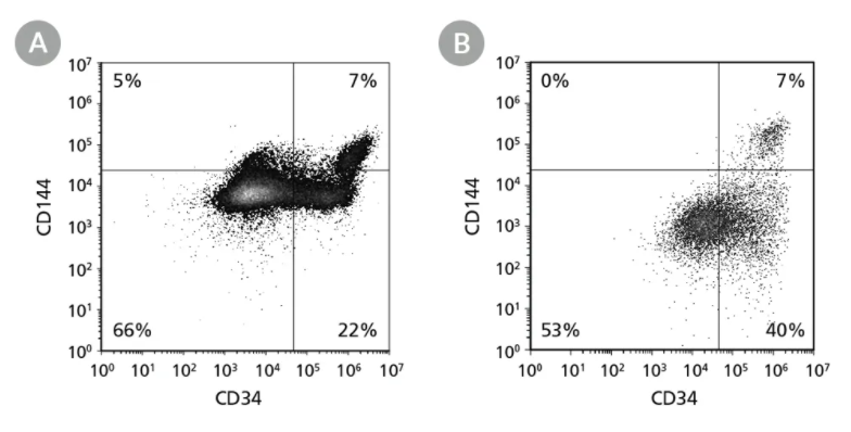
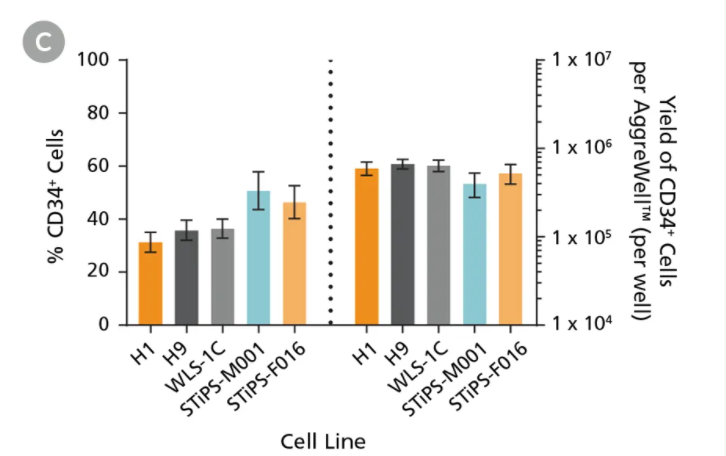
Figure 2. hPSCs Differentiate to CD34+ Hematopoietic Progenitor Cells After 12 Days of CultureHuman ES and iPS cells were induced to differentiate to CD34+ cells using the 12-day protocol shown in Figure 1. At the end of the culture period, cells were harvested, dissociated into a single-cell suspension, and analyzed by flow cytometry for CD34 and CD144 expression. Dead cells were excluded by light-scatter profile and DRAQ7™ staining. Representative flow cytometry plots for (A) ES (H1) cell-derived and (B) iPS (STiPS-M001) cell-derived cells analyzed on day 12 are shown. (C) The average frequency of viable CD34+ cells on day 12 (before CD34+ cell isolation) for two ES cell lines (H1 and H9) and three iPS cell lines (WLS-1C, STiPS-M001, and STiPS-F016) ranged between 31% and 51%. The average yield of CD34+ cells produced per well of a 6-well AggreWell™400 plate ranged between 4.0 x 10⁵ and 6.7 x 10⁵. Data are shown as mean ± SEM (n = 9 - 35).
Figure 3. NK Cell Generation ProtocolhPSC-derived CD34+ cells are seeded in StemSpan™ Lymphoid Progenitor Expansion Medium on plates coated with StemSpan™ Lymphoid Differentiation Coating Material. On day 14, cells at the lymphoid progenitor stage are harvested and reseeded in StemSpan™ NK Cell Differentiation Medium onto non-coated plates for further differentiation to NK cells.
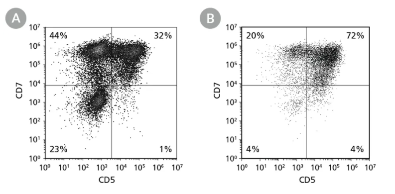
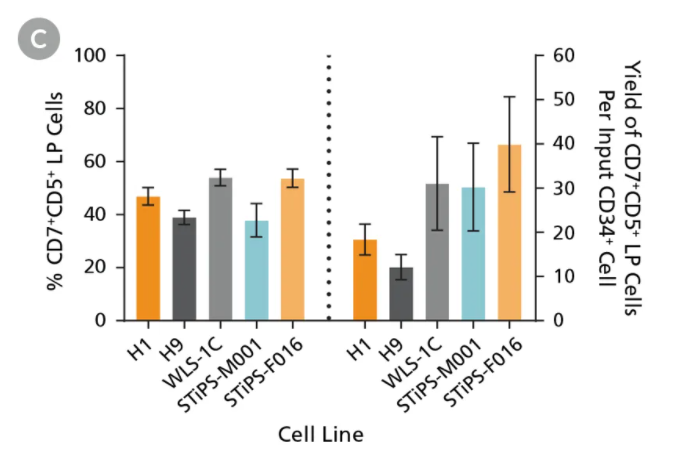
Figure 4. hPSC-Derived CD34+ Cells Differentiate to CD5+CD7+ Lymphoid Progenitor Cells Over 14 Days of CulturehPSC-derived CD34+ cells were cultured for 14 days in StemSpan™ Lymphoid Progenitor Expansion Medium on plates coated with StemSpan™ Lymphoid Differentiation Coating Material (Figure 2). Cells were harvested and analyzed for CD7 and CD5 expression by flow cytometry. Representative flow cytometry plots for (A) ES (H1) cell-derived and (B) iPS (STiPS-M001) cell-derived cells are shown. (C) The average frequency of viable CD7+CD5+ lymphoid progenitor cells on day 14 ranged between 38% and 54%, and the average yield of lymphoid progenitor cells produced per input hPSC-derived CD34+ cell was between 12 and 40. Data are shown as mean ± SEM (n = 8 - 32).
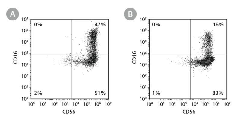
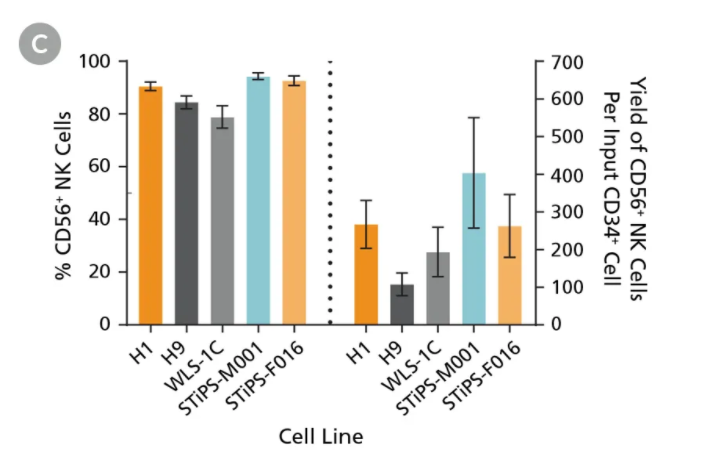
Figure 5. hPSC-Derived Lymphoid Progenitor Cells Differentiate to CD56+ NK Cells After 14 Days of CulturehPSC-derived lymphoid progenitor cells were cultured in StemSpan™ NK Cell Differentiation Medium on non-coated plates for 14 days (Figure 2). Cells were harvested and analyzed for expression of CD56 and CD16 by flow cytometry. Representative flow cytometry plots are shown for both (A) ES (H1) cell-derived and (B) iPS (STiPS-M001) cell-derived cells. (C) After 28 days of culture, the average frequency of viable CD56+ NK cells from hPSC-derived CD34+ cells ranged between 79% and 94%. The average yield of CD56+ cells produced per hPSC-derived CD34+ cell was between 108 and 404. Data are shown as mean ± SEM (n = 7 - 18).
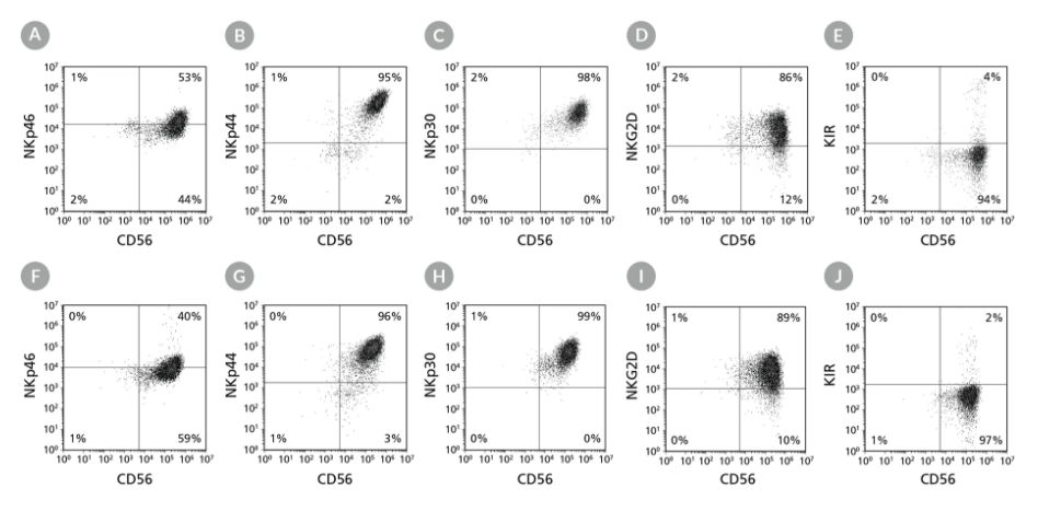
Figure 6. Cell Surface Marker Expression on PSC-Derived CD56+ NK Cells After 28 Days of CulturehPSC-derived CD34+ cells were cultured in StemSpan™ Lymphoid Progenitor Expansion Medium on plates coated with StemSpan™ Lymphoid Differentiation Coating Material for 14 days, followed by 14 days of culture in StemSpan™ NK Cell Differentiation Medium on non-coated plates to generate CD56+ NK cells (Figure 2). Cells were harvested and analyzed for CD56, NKp46, NKp44, NKp30, NKG2D, and KIR expression by flow cytometry. Dead cells were excluded by light-scatter profile and DRAQ7™ staining. Data shown are from representative cultures initiated with (A-E) ES (H1) cells or (F-J) iPS (STiPS-M001) cells.
Figure 7. Cultured NK Cells Exhibit Cytotoxicity Toward K562 Cell LinehPSC-derived CD56+ NK cells were co-cultured with calcein AM (CAM)-labeled K562 cells at a ratio of 2.5:1 for 4 hours. Isolated peripheral blood (PB) NK cells and monocytes were also co-cultured with labeled K562 cells as positive and negative controls, respectively. Before the co-culture, frozen PB NK cells were thawed and cultured overnight with StemSpan™ NK Cell Differentiation Supplement and StemSpan™ SFEM II, while PB monocytes were cultured overnight in SFEM II only. To measure maximum release, the labeled K562 cells were treated with 1% Triton™ X-100. Culture supernatants were assessed for fluorescence released by dead cells after 4 hours using a SpectraMax® microplate reader (excitation 485 nm/emission 520 nm). The % specific lysis was calculated as follows: [(test release - spontaneous release) / (maximum release - spontaneous release)] x 100%. The average specific lysis by hPSC- derived NK cells ranged between 51% and 75% as compared to 62% specific lysis by PB NK cells. Cultures of ES (H1 and H9) and iPS (WLS-1C, STiPS-M001, and STiPS-F016) cells are shown. Data are shown as mean ± SEM (n = 3 - 7).
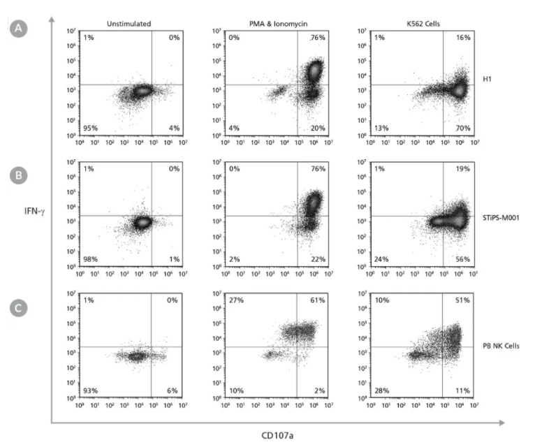
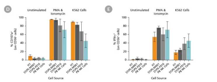
Figure 8. Cultured hPSC-Derived CD56+ NK Cells Are Induced to Express Surface CD107a and Produce IFN-γ After Co-Culture with K562 Target Cells or Stimulation with PMA and IonomycinhPSC-derived NK cells were obtained using STEMdiff™ NK Cell Kit as described. hPSC-derived CD56+ NK cells were left unstimulated or were stimulated with either phorbol 12-myristate 13-acetate (PMA) and ionomycin or with K562 cells at a ratio of 1:1 effector to target cells. CD107a antibody and stimulation factors were added to each well. After one hour, monensin and brefeldin A were added to each well to inhibit protein transport. After a total incubation time of 4 hours at 37°C, cells were washed and stained with Zombie NIR™ Fixable Viability Kit and CD56. Cells were then fixed, permeabilized, and stained for IFN-γ. Surface CD107a and intracellular IFN-γ expression were assessed using flow cytometry. Representative samples are gated on CD56. (A) Representative unstimulated, PMA & ionomycin- stimulated, and K562-stimulated ES (H1) cell-derived NK cell samples. (B) Representative unstimulated, PMA & ionomycin-stimulated, and K562-stimulated iPS (STiPS-M001) cell-derived NK cell samples. (C) Representative unstimulated, PMA & ionomycin-stimulated, and K562-stimulated PB NK cell samples. The PB NK cell sample was thawed and cultured overnight in StemSpan™ SFEM II supplemented with IL-2 prior to this assay. (D) Summary of CD107a and (E) IFN-γ expression results by NK cells. Upon stimulation, hPSC-derived CD56+ NK cells are able to degranulate, as shown by surface expression of CD107a (average range: 82 - 96% for PMA & ionomycin stimulation and 68 - 90% for K562 stimulation) and secrete IFN-γ (54 - 76% and 18 - 39% for PMA & ionomycin and K562 stimulation, respectively). Data are shown as mean ± SEM (n = 2 - 4 ). |